
Abstract
Background The prognosis and treatment outcomes for patients with rectal cancer are critically dependent on an accurate and comprehensive preoperative evaluation.Three-dimensional endorectal ultrasound (3D-ERUS) has demonstrated high accuracy in the T staging of rectal cancer. Thus, we aimed to develop a computer-aided diagnosis (CAD) tool using a deep learning model for the preoperative T-staging of rectal cancer with 3D-ERUS.
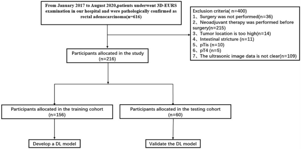
Methods We retrospectively analyzed the data of 216 rectal cancer patients who underwent 3D-ERUS. The patients were randomly assigned to a training cohort (n=156) or a testing cohort (n=60). Radiologists interpreted the 3D-ERUS images of the testing cohort with and without the CAD tool. The diagnostic performance of the CAD tool and its impact on the radiologists’ interpretations were evaluated.
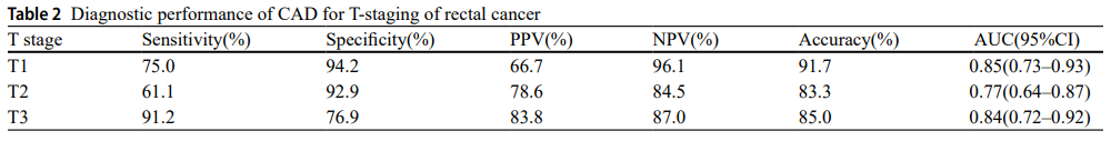
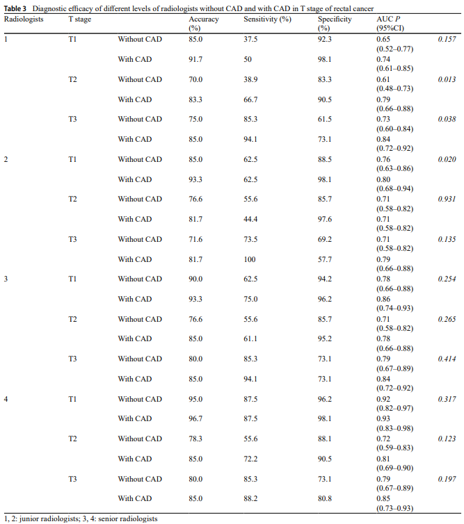
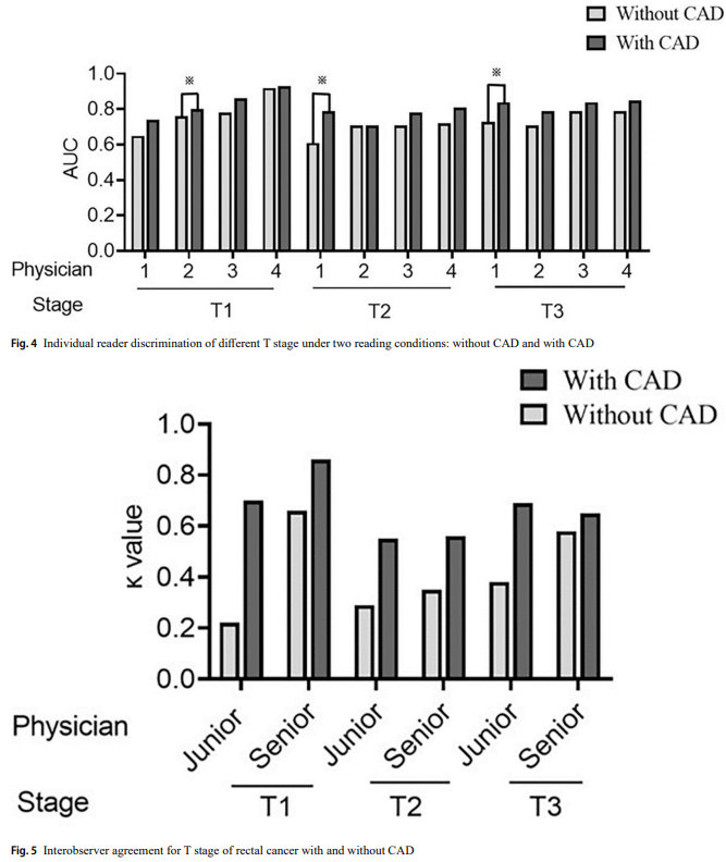
Results The CAD tool demonstrated high diagnostic efficacy for rectal cancer tumors of all T stages, with the best diagnostic performance achieved for T1-stage tumors (AUC, 0.85; 95% CI, 0.73–0.93). With assistance from the CAD tool, the AUC for T1 tumors improved from 0.76 (95% CI, 0.63–0.86) to 0.80 (95% CI, 0.68–0.94) (P=0.020) for junior radiologist 2. For junior radiologist 1, the AUC improved from 0.61 (95% CI, 0.48–0.73) to 0.79 (95% CI, 0.66–0.88) (P=0.013) for T2 tumors and from 0.73 (95% CI, 0.60–0.84) to 0.84 (95% CI, 0.72–0.92) (P=0.038) for T3 tumors. The diagnostic consistency (κ value) also improved from 0.31 to 0.64 (P=0.005) for the junior radiologists and from 0.52 to 0.66 (P=0.005) for the senior radiologists.
Conclusion A CAD tool utilizing a deep learning model based on 3D-ERUS images showed strong performance in T staging rectal cancer. This tool could improve the performance of and consistency between radiologists in preoperatively assessing rectal cancer patients.
Framework
Experiment
Conclusion
The results of this study demonstrated that an AI-based CAD tool can enhance radiologists’ performance and agreement in T-staging rectal cancer tumors. The aim of this study was to develop an automated diagnostic model for T staging rectal cancer whose performance is comparable to or surpasses that of experienced radiologists. Integrating medicine and engineering in a DL model yielded promising results and provides support for advancing CAD tools toward clinical implementation. Future prospective studies will be essential for assessing the impact of CAD systems on accurately and comprehensively assessing rectal cancer patients.
- hcp@sysu.edu.cn
- 广州市广州大学城外环东路132号

- Projects
- Computer Vision
- Multimodal
- Robotics
- Links
- Git-Lab